Publications

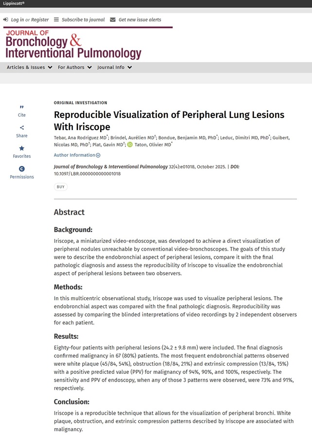
Reproducible Visualization of Peripheral Lung Lesions With Iriscope
This multicentre observational study performed in Hôpital Erasme (Belgium) and CHU Toulouse (France) evaluated Iriscope in 84 patients (mean lesion size ≈ 24 mm) with peripheral lung lesions. Iriscope revealed three malignancy‑linked patterns—white plaque, obstruction and extrinsic compression—achieving 94–100 % PPV and 73 % overall sensitivity. Strong inter‑observer agreement confirmed reproducibility. These findings underline Iriscope’s capacity to characterise peripheral lesions and steer precise, ultrasound‑complementing sampling.

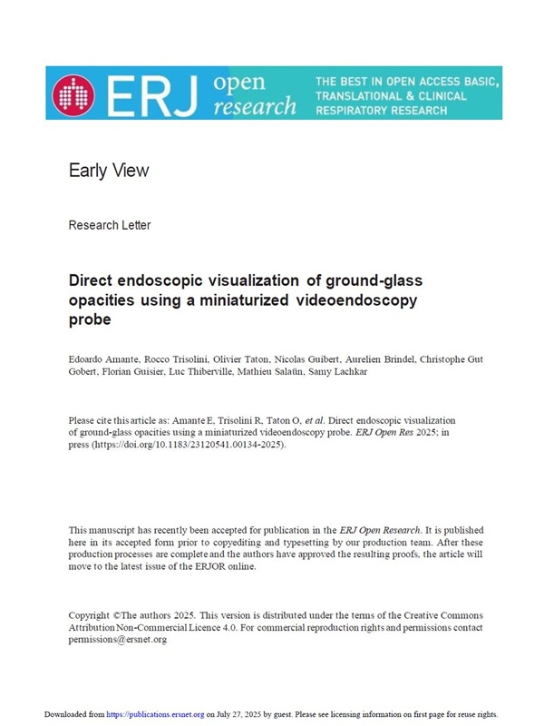
Direct endoscopic visualization of ground-glass opacities using a miniaturized videoendoscopy probe
This multicentre retrospective study performed in CHU Rouen, CHU Toulouse, CHU Brest (Fance) and Hôpital Erasme (Belgium) assessed a 1.3 mm Iriscope probe in 63 pulmonary ground‑glass opacities (30 pure, 33 mixed). Iriscope visualised every lesion and delivered 100 % specificity and positive predictive value, with sensitivities of 68 % for pure and 70 % for mixed GGOs when predicting malignancy. Notably, it revealed tumoral features in 50 % of cancers that were invisible on r‑EBUS, and no complications occurred. These findings highlight Iriscope’s ability to refine GGO diagnosis and guide precise sampling when conventional ultrasound is inconclusive.

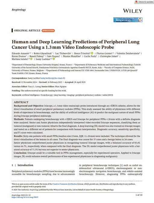
Human and Deep Learning Predictions of Peripheral Lung Cancer Using a 1.3 mm Video Endoscopic Probe
This single‑centre study by Edoardo Amante et al., from Dr Samy Lachkar’s team, assessed a 1.3 mm Iriscope probe introduced in 61 patients with peripheral pulmonary nodules ≤20 mm. The probe enabled direct visualisation of every lesion and, when images were read by senior bronchoscopists, delivered a balanced accuracy of 85 % for malignancy prediction. Junior physicians were less accurate, but a deep‑learning model trained on 62 000 Iriscope frames outperformed them (balanced accuracy ≈72 %), narrowing the expertise gap. These findings confirm the feasibility of real‑time peripheral endoscopy and suggest that combining Iriscope imaging with AI support could sharpen diagnostic confidence and guide precise sampling of small lung nodules.

Direct endoscopic visualization of small peripheral lung nodules using a miniaturized videoendoscopy probe
This retrospective study by Samy Lachkar et al. examines the use of the Iriscope probe combined with radial endobronchial ultrasound (r-EBUS) to directly visualize small peripheral pulmonary nodules. In 23 patients, the technique allowed direct endoscopic visualization in the majority of cases, improving diagnosis. Analysis of Iriscope images showed a high positive predictive value for cancer diagnosis (93%). The study highlights the potential of this approach to improve diagnostic yield and guide therapeutic strategy, despite some limitations. The authors suggest that this simple technique could optimize the accuracy of sampling and diagnosis of pulmonary nodules.

Feasibility and impact on diagnosis of peripheral pulmonary lesions under real-time direct vision by Iriscope
This prospective study from Borja Recalde-Zamacona et al. evaluates the effectiveness and diagnostic impact of the Iriscope in the real-time visualization of peripheral pulmonary lesions (PPL). Combined with radial endobronchial ultrasound (rEBUS) and/or electromagnetic navigation bronchoscopy (ENB), Iriscope allowed direct visualization of 57.1% of PPLs, with a high positive predictive value for the diagnosis of malignant lesions (92.5%). The results suggest that the Iriscope is a promising and complementary technique, but larger studies are needed to confirm its impact on overall diagnostic yield and to identify predictive patterns. Finally, the safety of the procedure using the Iriscope was confirmed.

Emphysema Seen through a Mini-Camera during an Electromagnetic Navigation Bronchoscopy Guided by Cone-Beam Computed Tomography
This case report by Prof. Dimitri Leduc's team details the visualization of emphysema using Iriscope during an electromagnetic navigation bronchoscopy (ENB) guided by cone-beam computed tomography (CBCT). The technique provided detailed imaging of lung lesions, offering valuable insights into the condition. The findings highlight the potential of this approach in enhancing the diagnostic process for pulmonary diseases.

Direct visualization of peripheral lung nodules with Iriscope
A multicenter study by Olivier Taton et al. evaluated the efficacy and safety of Iriscope for the direct visualization of peripheral pulmonary nodules. In 96 patients, Iriscope effectively visualized the peripheral bronchi with a high success rate despite the presence of blood. Various endobronchial patterns were observed and compared to the final pathological diagnosis, revealing good correlation in the majority of cases. The inter-observer concordance rate was significant, demonstrating the reproducibility of the method. The study highlights the usefulness of the Iriscope in diagnosing peripheral pulmonary lesions.

New ultrathin videoprobe for peripheral pulmonary lesion
A prospective monocentric study from Olivier Taton et al. compared the efficacy of Iriscope to radial endobronchial ultrasound (R-EBUS) for visualizing peripheral pulmonary lesions. The results suggest non-inferiority of the Iriscope compared to R-EBUS, with improved diagnostic yield when combining both techniques. The study included patients with lesions ranging from 20 to 50 mm, randomized into three groups: Iriscope alone, R-EBUS alone, and both combined. The Iriscope demonstrated high sensitivity, specificity, PPV, and NPV, especially when combined with R-EBUS.